Oak Hill Dogs Killed After Having Brain Matter Suctioned Out
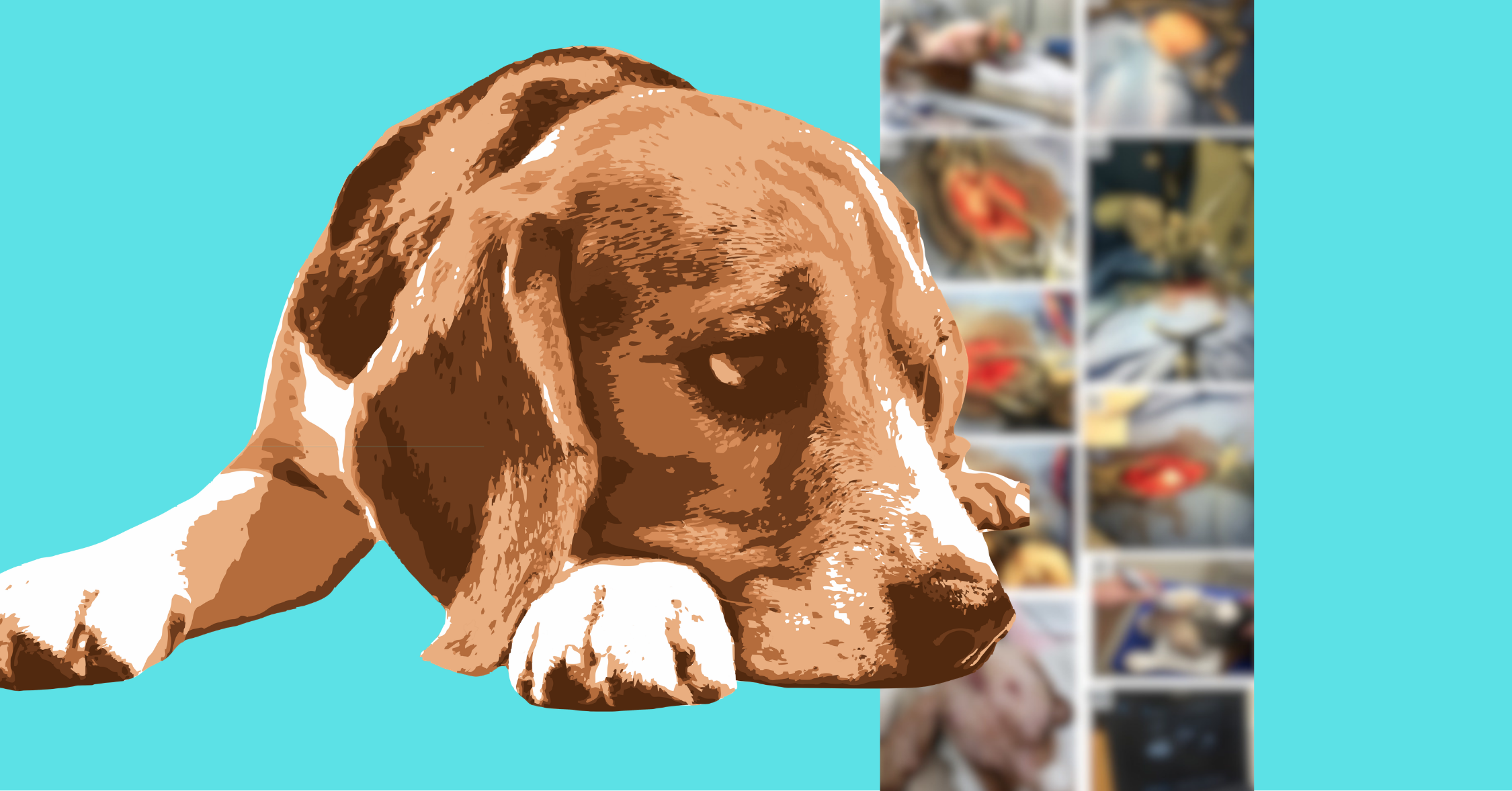
Four young male dogs underwent brain surgery, spinal fluid draws, and other horrors before being killed in a federally funded brain cancer experiment run jointly by North Carolina State University and the University of North Carolina at Chapel Hill (UNC).
These “purpose-bred” beagles—all between one and two years old, and identified only as CP01, CP02, CP03, and CP04—came from Oak Hill Genetics, the country’s second-largest breeder of dogs for experimentation.

The paper resulting from this experiment recounts what the dogs endured in highly technical terms. Here is what it describes, plainly:
Researchers started by cutting out a piece of each dog’s neck.
From the back of each dog’s neck, researchers removed a full-thickness skin sample. Cells from those samples were then manipulated in the lab, converted into a different type of cell and engineered with two methods of attacking cancer. One was designed to kill cancer cells directly; the other required a drug, administered to the dogs weeks later, to become active.

Next, researchers cut open the dogs’ skulls and suctioned out parts of their brains.
When surgeons remove a brain tumor from a human patient, they leave a hole behind. Researchers wanted to see whether the manipulated cells from the dogs’ own neck tissue would survive in a hole like that. So they made one.
They cut away a piece of each dog’s skull, opened the tough membrane underneath, and used suction to remove a square of healthy brain along with the tissue below it.

CP01’s and CP02’s holes were left empty, but researchers deposited into CP03’s and CP04’s holes a surgical gel containing the cells grown from their own neck skin.
Afterward, they did not sew the membrane back together or replace the piece of skull. Instead, they merely pulled the membrane closed by hand and covered it with a synthetic patch.
Two of the dogs had hardware implanted in their skulls.

Researchers drilled a second hole in the skulls of CP01 and CP02, then ran a tube down toward one of the fluid-filled chambers deep inside the brain and attached it to a small plastic dome that they set into the hole in the skull. A dose of cells went into the dome before the chewing muscle, the tissue beneath the skin, and the skin itself were closed over the top of it.
Over the next two months, researchers pushed a needle through the skin and through that muscle into the dome twice more to deliver two further doses of cells.
In neither dog’s brain did the tube end up precisely where researchers had aimed.
In CP01, it ran past the chamber; in CP02, it sat just outside it. The researchers’ excuses? The bite plates used to guide the surgery did not fit the dogs’ teeth properly, and dog brain chambers are small.
Each dog was given a drug carrying the FDA’s most serious boxed warning.
The experimental cells were designed to attack cancer only in part on their own. The other half of that design had to be switched on by a drug the dogs were forced to ingest: valganciclovir.

The drug carries an FDA boxed warning for several serious risks, including damage to blood cells and reproductive toxicity.
Both would become troubling findings in the experiment.
The months between surgery and death were agonizing.
- The dogs were repeatedly anesthetized for scans and other procedures, and they had their blood drawn over and over.
- Researchers pierced the bases of their skulls with needles to draw spinal fluid. Tissue examinations later revealed bleeding in the spinal cords of all four dogs, which the researchers attributed to spinal fluid collection around the time of death.
- Researchers forced tubes into their bladders to collect urine.
- Three of the dogs came out of brain surgery unable to see properly on one side, and all four dogs experienced difficulty controlling their legs on that same side. Researchers attributed both issues to the brain tissue they had suctioned out rather than to the experimental cells—which is to say the damage came from the holes the researchers had deliberately made in the dogs’ brains.
- All four dogs experienced repeated crashes of white blood cells that fight infections.
- All four dogs’ testicles were damaged. Evidence of testicular degeneration and abnormalities in sperm production turned up first in CP01 and CP03, when their bodies were examined after death. Then, in a supposed effort to identify the cause, researchers took one testicle each from CP02 and CP04, the two dogs still alive. Researchers ultimately blamed the drug valganciclovir for the reproductive damage—and, despite the clear FDA boxed warning about reproductive toxicity, they characterized the dogs’ testicular damage as surprising and unexpected.
- Inside the dogs’ heads, the tissue around the implantation sites (around the gel in two dogs, around the hardware in the other two) were inflamed and scarred, with nerve fibers breaking down. In CP01 and CP02, the membranes around the holes in their brains were thickened and inflamed, as well.
At the end of the study, each dog was killed and immediately dissected.
To justify all of this, researchers framed the dogs as a bridge to human patients—but that bridge went nowhere.
The paper is clear about why dogs were exploited. For one thing, dogs, like humans, can develop brain tumors spontaneously. But the four dogs chosen were “healthy” and tumor-free. Researchers’ justification was that the spontaneous-tumor similarity mattered not for this experiment, but for the efficacy studies they intended to run on more dogs in the future.
In other words, one of the stated reasons for cutting open these four dogs was a plan to cut open more in the future.
The authors also contended that a large animal was needed to bridge the gap between mouse studies and a first trial in human patients. Those who stood to gain directly by that gap closing appear only at the back of the paper, in the “conflicts of interest” section: Falcon Therapeutics, Inc. had licensed aspects of the technology under study from UNC, and two of the paper’s nine authors—both UNC faculty—held ownership interests in it. One was a co-founder who sat on its board of directors.
Though the paper promised that the results it conveyed “pave[d] the way for future human clinical trials,” Falcon Therapeutics filed for Chapter 7 bankruptcy and shut down 14 months after the paper’s publication.
Dogs were not the only animals harmed.
Before the dog experiments, researchers exploited an unspecified number of female, six-to-eight-week-old mice who had been bred to have compromised immune systems.
The mice all had an incision cut down the middle of their scalps and holes drilled in their skulls. Some then had dog skin cells—not even the real therapy, which researchers skipped to save time—injected straight into their brains. Three days later, they were killed by pumping saline and a preservative through their hearts, so their brains could be removed and examined.
The paper plainly says that “immune-depleted” mice cannot reveal what researchers needed to know about the experimental therapy—and researchers used this to justify moving on to dogs.
No one knows how many animals lost their lives for this research.
The number of mice brutalized and killed for this experiment remains unreported.
But we do now know about four individual canine victims:
CP01, CP02, CP03, and CP04 were bred to be exploited, shipped to a lab, taken apart in pieces, and killed after months of suffering—then reduced to technical jargon across the pages of a journal.
They were four dogs among the tens of thousands who research facilities are buying, holding, and using right now. We may be too late to save them, but, together, we can still save the others.